
Wildlife
“When people touch an animal, the animal touches their heart. And instantly, we’ve won them over to the conservation of that species.”
Steve Irwin
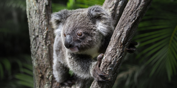
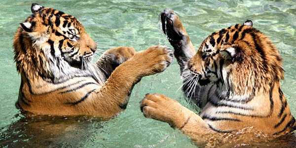
Our Animals
We just love animals – from the tiniest lizard to the tallest giraffe! Whether you are mad about mammals, think birds are brilliant or really like reptiles, this is for you.
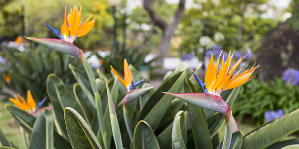
Zoo Flora
Alongside our amazing animals, the Zoo is home to a stunning array of native and exotic flora. You can enjoy browsing a selection of plants that call Australia Zoo home.
Top 10 Highlights
It was hard sticking to 10, but we’ve compiled a list of what we think are Australia Zoo’s Top 10 Highlights for our visitors!