Work Experience
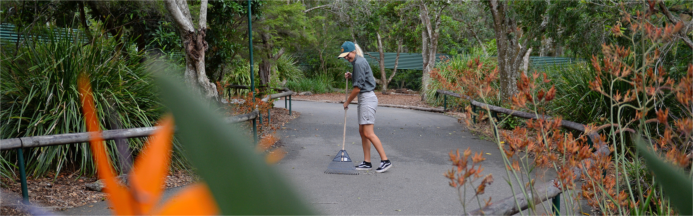
Introduction to Zoo Keeping participants will assist with a range of activities associated with the care of animals at Australia Zoo. Activities may include, but not be limited to, assisting with habitat cleans (raking, sweeping, weeding, scrubbing), animal observations and the preparation of diets and enrichment items. This program also incorporates educational workshops and presentations on relevant industry topics, as well as daily debrief/review sessions.
The departments that high school participants may assist with include Birds and Australasian Mammals.
It is important to note that, due to a range of operational, legislative and animal welfare considerations, participants should not have any expectation of direct animal interaction.
Applicants will be required to participate in an interview and selection process. Please note that no placements within the program can be guaranteed until after this interview process has been completed. Any associated travel, accommodation or school commitments should not be made until a confirmation of placement is made by Australia Zoo. Placement on all programs is at the discretion of Australia Zoo.
Please refer to placement and application dates below:
The placement dates available for 2024 are:
| Placement Dates From | Placement Dates To | Applications Open | Applications Close |
| Term One | |||
| Monday 18th March | Thursday the 21st March | Monday the 8th January | Monday the 5th February |
| Term Two | |||
| Monday 17th June | Thursday the 20th June | Monday the 8th April | Monday 6th May |
| Term Three | |||
| Monday the 9th September | Thursday the 12th September | Monday the 1st July | Monday the 29th July |
| Term Four | |||
| Monday the 18th November | Thursday the 21st November | Monday the 2nd September | Monday the 30th September |
Programs run from 9am-3pm each day
Ready to apply?
All applicants are required to complete all sections of the online Work Experience Application form which will include a picture, your current resume, references and proof of vaccinations. – Online Volunteer Application
Applications cannot be processed unless all information is provided. Once your application has been reviewed, you will notified as to whether you have progressed to the next stage, which will include an online interview.
Further information
The following information packs are available to download:
- Participant, parent and guardian information – download here
- Additional information for school coordinators – download here
We look forward to receiving your application. Please contact us if you have any further questions.